Mammalian Heart Dissection Guide Doc

Sheep Heart Dissection Lab Answers
. Share. Sheep have a four-chambered heart, just like humans.
By studying the anatomy of a sheep’s heart, you can learn about how your own heart pumps blood through your body and keeps you alive! Use this as a dissection guide complete enough for a high school lab, or just look at the labeled images to get an idea of what the heart looks like. If you do the dissection yourself, you will need a. Observation: External Anatomy Most heart diagrams show the left atrium and ventricle on the right side of the diagram. Imagine the heart in the body of a person facing you. The left side of their heart is on their left, but since you are facing them, it is on your right. Click for full-size pdf 1.
Identify the right and left sides of the heart. Look closely and on one side you will see a diagonal line of blood vessels that divide the heart.
The half that includes all of the apex (pointed end) of the heart is the left side. Confirm this by squeezing each half of the heart.
The left half will feel much firmer and more muscular than the right side. (The left side of the heart is stronger because it has to pump blood to the whole body. The right side only pumps blood to the lungs.) 2. Turn the heart so that the right side is on your right, as if it were in your body.
Examine the flaps of darker tissue on the top of the heart. These ear-like flaps are called auricles. Find the large opening at the top of the heart next to the right auricle. This is the opening to the superior vena cava, which brings blood from the top half of the body to the right atrium (the atria are the top chambers in the heart).
Stick a down this vessel. You should feel it open into the right atrium. Click for full-size pdf A little down and to the left of the superior vena cava there is another blood vessel opening. Insert your probe into this; it should also lead into the right atrium. This is the inferior vena cava, which brings blood from the lower tissues. You can also see another blood vessel next to the left auricle. This is a pulmonary vein that brings blood from the lungs into the left atrium.
Sticking straight up from the center of the heart is the largest blood vessel you will see. This is the aorta, which takes oxygenated blood from the left ventricle to the rest of the body (the ventricles are the lower chambers of the heart). The aorta branches into more than one artery right after it leaves the heart, so it may have more than one opening on your heart specimen.
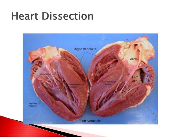
Look carefully at the openings and you should be able to see that they are connected to each other. Behind and to the left of the aorta there is another large vessel. This is the pulmonary artery which takes blood from the right ventricle to the lungs. Dissection: Internal Anatomy 1. Insert your or Click for full-size pdf into the superior vena cava and make an incision down through the wall of the right atrium and ventricle, as shown by the dotted line in the external heart picture. Pull the two sides apart and look for three flaps of membrane. These membranes form the tricuspid valve between the right atrium and the right ventricle.
The membranes are connected to flaps of muscle called the papillary muscles by tendons called the chordae tendinae or “heartstrings.” This valve allows blood to enter the ventricle from the atrium, but prevents backflow from the ventricle into the atrium. Insert your probe into the pulmonary artery and see it come through to the right ventricle. Make an incision down through this artery and look inside it for three small membranous pockets. These form the pulmonary semilunar valve which prevents blood from flowing back into the right ventricle. Insert your dissecting scissors or scalpel into the left auricle at the base Click for full-size pdf of the aorta and make an incision down through the wall of the left atrium and ventricle, as shown by the dotted line in the external heart picture. Locate the mitral valve (or bicuspid valve) between the left atrium and ventricle. This will have two flaps of membrane connected to papillary muscles by tendons.
Insert a probe into the aorta and observe where it connects to the left ventricle. Make an incision up through the aorta and examine the inside carefully for three small membranous pockets. These form the aortic semilunar valve which prevents blood from flowing back into the left ventricle. Blood Flow Now consider all the parts you’ve found and how the blood flows through them.
Draw a diagram of the heart and use red and blue arrows to show the flow of blood: → deoxygenated blood → oxygenated blood Blood from the tissues → superior and inferior vena cava → right atrium → tricuspid valve → right ventricle → pulmonary semilunar valve → pulmonary artery → lungs → pulmonary veins → left atrium → bicuspid (mitral) valve → left ventricle → aortic semilunar valve → aorta → body tissue. Label the Parts of a Sheep Heart Print out these diagrams and fill in the labels to test your knowledge of sheep heart anatomy. External anatomy: (.pdf).
Internal anatomy: (.pdf). Internal anatomy: (.pdf) See our other free dissection guides with photos and printable PDFs.
Lost your user manual? At eSpares we work hard to maintain a library of manufacturer-approved guides and instructions for.

We're always expanding our collection, so if the manual you need isn't here, let us know and we'll do our best to find it for you. Are you looking for? You will need Adobe Acrobat Reader® installed on your computer in order to display these files. If you do not already have such on your computer, search on search engine to download a free copy of Acrobat Reader®. If you are looking for that will help you solve your problem, then our online library is for you.
There are many of Answerkey guides, and many ebooks related with. But very firstly read carefuly of privacy policy of statement.